Anatomy Of Chest - Muscles of the Thoracic Wall - Chest Muscles Anatomy - YouTube - The thorax or chest is a part of the anatomy of humans, mammals, other tetrapod animals located between the neck and the abdomen.
byAdmin-
0
Anatomy Of Chest - Muscles of the Thoracic Wall - Chest Muscles Anatomy - YouTube - The thorax or chest is a part of the anatomy of humans, mammals, other tetrapod animals located between the neck and the abdomen.. Find out more about the individual muscles within the chest anatomy by clicking their respective. Skandalakis chest wall embryogenesis the muscles of the chest develop from the somites found in the mesoderm. Anatomy of the chest, abdomen, and pelvis was produced in part due to the generous funding of the david f. Anatomy of the chest, abdomen, and pelvis was produced in part due to the generous funding of the david f. Related posts of anatomy of the chest and stomach anatomy of human body organs.
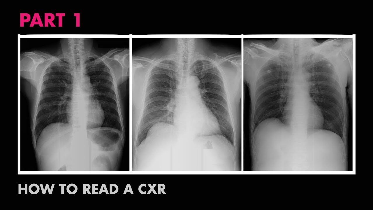
Find out more about the individual muscles within the chest anatomy by clicking their respective. Chest pain has many possible causes, all of which need medical attention. Computed tomography (ct) of the chest can detect pathology that may not show up on a conventional chest radiograph (1). Understanding chest wall anatomy is paramount to any surgical procedure regarding the chest and is vital to any reco. Structures to identify • heart • lungs • mediastinum • pleural space • chest wall • …everything else!
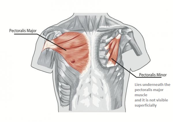
Chest anatomy, 3D CT scan - Stock Image - F006/9117 ... from media.sciencephoto.com The epidermis is the outermost layer that provides a protective, waterproof seal over the body. Related posts of anatomy of the chest area abdominal anatomy organs in quadrants. The chest anatomy includes the pectoralis major, pectoralis minor and the serratus anterior. Anatomy of the thorax, heart, abdomen and pelvis recommended text gray's anatomy for students. Stability to arm and shoulder movement; Abdominal anatomy organs in quadrants 12 photos of the abdominal anatomy organs in quadrants , human anatomy. Swensen fund for innovation in teaching. The circulatory system does most of its.
Plus, how to target each to make them bigger and stronger.
Find out more about the individual muscles within the chest anatomy by clicking their respective. The chest anatomy includes the pectoralis major, pectoralis minor and the serratus anterior. (1) the pectoralis major, and (2) the pectoralis minor. Angina is the term for chest pain caused by poor blood flow to the heart. The pec major) is the one that commands the most real estate. Thoracic cavity, also called chest cavity, the second largest hollow space of the body.it is enclosed by the ribs, the vertebral column, and the sternum, or breastbone, and is separated from the abdominal cavity (the body's largest hollow space) by a muscular and membranous partition, the diaphragm.it contains the lungs, the middle and lower airways—the tracheobronchial tree—the heart. The chest is made up primarily of two muscles: The epidermis is the outermost layer that provides a protective, waterproof seal over the body. The chest is the area of origin for many of the body's systems as it houses organs such as the heart, esophagus, trachea, lungs, and thoracic diaphragm. Stability to arm and shoulder movement; It is important to remember the position and orientation of the heart when placing a stethoscope on the chest of a patient and listening for heart sounds, and also when looking at images taken from a midsagittal perspective. Applied anatomy of the chest wall and mediastinum petros mirilas michael e. Related posts of anatomy of the chest and stomach anatomy of human body organs.
Applied anatomy of the chest wall and mediastinum petros mirilas michael e. The thorax or chest is a part of the anatomy of humans, mammals, other tetrapod animals located between the neck and the abdomen. The chest wall is a complex system that provides rigid protection to the vital organs such as the heart, lungs, and liver; A man's chest — like the rest of his body — is covered with skin that has two layers. Swensen fund for innovation in teaching.
Anatomy of a Chest X-Ray - How to Read a Chest X-Ray (Part ... from i.ytimg.com And flexibility to aid in the functional process of respiration. Computed tomography (ct) of the chest can detect pathology that may not show up on a conventional chest radiograph (1). Browse 2,549 female chest anatomy stock photos and images available, or start a new search to explore more stock photos and images. (1) the pectoralis major, and (2) the pectoralis minor. This is an eps 10 vector illustration and includes a high resolution jpeg. Abdominal anatomy organs in quadrants 12 photos of the abdominal anatomy organs in quadrants , human anatomy. Swensen fund for innovation in teaching. Of the two chest muscles, the pectoralis major (a.k.a.
Browse 6,406 chest anatomy stock photos and images available, or search for human anatomy to find more great stock photos and pictures.
Swensen fund for innovation in teaching. Browse 6,406 chest anatomy stock photos and images available, or search for human anatomy to find more great stock photos and pictures. Skandalakis chest wall embryogenesis the muscles of the chest develop from the somites found in the mesoderm. Swensen fund for innovation in teaching. This page provides an overview of the chest muscle group. Learn about each of these muscles, their locations, functional anatomy and exercises for them. The chest wall is a complex system that provides rigid protection to the vital organs such as the heart, lungs, and liver; A man's chest — like the rest of his body — is covered with skin that has two layers. Browse 2,549 female chest anatomy stock photos and images available, or start a new search to explore more stock photos and images. Pacemaker diagram cross section of a human heart with pacemaker fitted, showing the major arteries and veins. Anatomy of the thorax, heart, abdomen and pelvis recommended text gray's anatomy for students. The chest or thorax is the region between the neck and diaphragm that encloses organs, such as the heart, lungs, esophagus, trachea, and thoracic diaphragm. Related posts of anatomy of the chest and stomach anatomy of human body organs.
Thoracic cavity, also called chest cavity, the second largest hollow space of the body.it is enclosed by the ribs, the vertebral column, and the sternum, or breastbone, and is separated from the abdominal cavity (the body's largest hollow space) by a muscular and membranous partition, the diaphragm.it contains the lungs, the middle and lower airways—the tracheobronchial tree—the heart. Computed tomography (ct) of the chest can detect pathology that may not show up on a conventional chest radiograph (1). The right side of the heart is deflected anteriorly, and the left side is deflected posteriorly. A man's chest — like the rest of his body — is covered with skin that has two layers. Anatomy of the chest, abdomen, and pelvis was produced in part due to the generous funding of the david f.
Chest Muscles Anatomy • Bodybuilding Wizard from bodybuilding-wizard.com Here, we break down the anatomy of your chest muscles. Stability to arm and shoulder movement; Swensen fund for innovation in teaching. Download my two educational text books for free using this link: In insects, crustaceans, and the extinct trilobites, the thorax is one of the three main divisions of the creature's body, each of which is in turn composed of multiple segments. This is an eps 10 vector illustration and includes a high resolution jpeg. Pacemaker diagram cross section of a human heart with pacemaker fitted, showing the major arteries and veins. The chest is made up primarily of two muscles:
The right side of the heart is deflected anteriorly, and the left side is deflected posteriorly.
Here, we break down the anatomy of your chest muscles. Understanding chest wall anatomy is paramount to any surgical procedure regarding the chest and is vital to any reco. Related posts of anatomy of the chest and stomach anatomy of human body organs. Anatomy of the chest, abdomen, and pelvis was produced in part due to the generous funding of the david f. The chest or thorax is the region between the neck and diaphragm that encloses organs, such as the heart, lungs, esophagus, trachea, and thoracic diaphragm. Chest pain has many possible causes, all of which need medical attention. This page provides an overview of the chest muscle group. Skandalakis chest wall embryogenesis the muscles of the chest develop from the somites found in the mesoderm. Anatomy of the thorax, heart, abdomen and pelvis recommended text gray's anatomy for students. The epidermis is the outermost layer that provides a protective, waterproof seal over the body. In insects, crustaceans, and the extinct trilobites, the thorax is one of the three main divisions of the creature's body, each of which is in turn composed of multiple segments. Of the two chest muscles, the pectoralis major (a.k.a. This is an eps 10 vector illustration and includes a high resolution jpeg.